Our cells are the building blocks of our bodies. They operate and function, provide the structure for our bodies, take in nutrients and do their specifed roles for our body, but they aren’t immortal. These cells must continually be replicated and be able to divide, to ensure our body can fully operate. That’s why cell replication is so important. Without it, we wouldn’t have any growth and repair capabilities thus it’s a vital procedure that must take place.
As already mentioned, our cells are the basic units of our bodies. Think of any part of your body; it’s made from one type of a cell or another. So replicating these cells are important. The actual act of replication occurs in the cell cycle. There are 5 main processes which occur in the cycle: G1, S, G2, M phase and Cytokenesis. In G1 the cell grows physically. New organelles are created and then the events of the S phase occur. S phase is where all the DNA replication of the cell cycle takes place. The actual processes of DNA replication have already been covered previously so be sure to check that out. After the S phase, the cell goes through G2 in which the cell undergoes further growth and protein synthesis occurs. All the steps mentioned above, all constitute the interphase portion of the cell cycle. This phase is where the cell mostly stays the duration of it’s time. It’s estimated that the cell spends approximately 90% of its time in this phase. The other 10% come from the mitosis section of the cell cycle. However, in some cases a cell can go through another stage completely, known as the G0 phase. Also known as the resting state, many cells spend their time in this phase either for rest or to perform assigned duties.
Moving on, the mitosis stage follows the G2 phase. It can be split up to many different sections: Prophase, prometaphase, metaphase, anaphase, telophase and finally (not strictly speaking a stage in mitosis but still important for the replication process) cytokinesis. We will cover all these steps in detail: what they do and why they are important.
Prophase:
In prophase, centrioles and the spindle fibers develop and chromatin fibres condense and shorten into chromosomes. Microtubules extend from the end poles of cells to the centre of the cells.
Prometaphase:
In this phase, the nuclear envelope dissolves and spindle fibres attach to kinetochore- which are located on the centromeres.
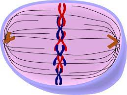
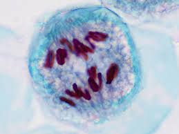
Metaphase:
The already replicated chromosomes line up at the equator of the cell (the metaphase plate). This is where most mitotic errors occur, as it determines the separation of sister chromatids.
Anaphase:
Following on from the chromosomes lining up along the metaphase plate, the spindle fibres retract and chromatids are separated moving to either poles of the cell. Spindle fibres from each centrosome pull the fibres with equal force in opposite directions. This is what allows the arms to separate and move towards the opposite poles.
Telophase:
The final section of mitosis is telophase. This is where the nuclear envelope reforms and the cytoplasm begins to cleave. The chromosomes are back inside the nucleus, but separated in daughter cells.
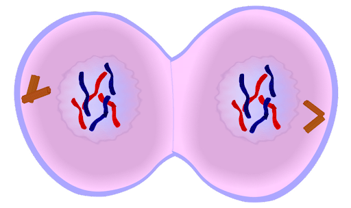
Cytokinesis:
The final phase of the cell cycle is cytokinesis. In this step, the cytoplasm cleaves even more resulting in the formation of 2 daughter cells. The cell membranes form in the individual daughter cells, separating the cells and finishing off mitosis. The cytokinesis stage is believed to actually begin from anaphase, where actin and myosin help with the cleavage formation, beginning the separation process.
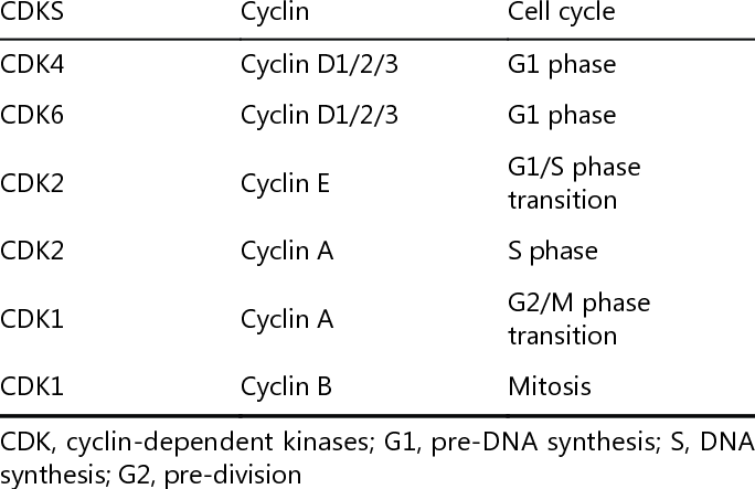
However despite the steps noted above, errors still occur. That’s were checkpoints come in. There are 3 checkpoints in the cell cycle: G1, G2 and M checkpoints. The G1 checkpoint checks to see if all the organelles have replicated and if there is enough proteins for DNA replication to go ahead. The G2 checkpoint checks to make sure that all the DNA is replicated in preparation for mitosis. Now the mitotic checkpoint occurs after metaphase to allow anaphase to proceed. It checks if all the chromosomes are on the metaphase plate and that they are all connected to kinetchores. They do this via signal proteins also known as Cdk proteins, which form complexes with cyclin proteins to check whether a cell is going through the right stages during the cell cycle. Cyclins are not always present during the cell cycle – and different cyclins get degraded at different stages of the cell cycle, depending on what stage stimulates their increase in concentration. This therefore means that different Cdks are active at different stages.
A table of stages, their cdk and cyclin complexes are shown below.
Some types of cell cycle errors can unfortunately get past the checkpoints and not get detected. Let’s take for example when incorrect cytokinesis occurs: this may be an error due to a lack of actin and myosin, which occurs due to medical conditions such as muscular dystrophy. There is no cell cycle checkpoint to check whether correct cytokinesis occurs: the only checkpoint in metaphase is to check if all chromosomes are attached to spindle fibres. Hence these issues would not be detected, and cause binucleate cells, for instance.
Some errors also occur by the incorrect placement of chromosomes on the metaphase plate. This is known as mitotic nondisjunction, caused by improper segregation. It may introduce new cell lines, which causes a type of variety known as mosaicism. Mosaicism is when you have more than 2 types of cell lines in your body. During anaphase, normally each homologous arm is pulled to the opposite pole of the cell, causing an even amount of chromosomes in each daughter cell. Sometimes, however, both arms of a chromosome go and split off at one pole and none goes to the other pole. This causes 3 arms of each chromosome during zygote fertilisation in one cell, also known as a trisomy. The other cell will only have 1 arm of a chromosome, also known as a monosomy. Bear in mind that most monosomies are lethal, with Monosomy 45 (Turner’s Syndrome) being the only monosomy that is compatible with life.
Nondisjunction can cause a variety of errors, which lead to diseases – some having major defects to a patient’s health. Examples of common diseases due to nondisjunction are stated below.
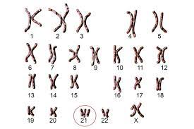
- Down’s Syndrome
Trisomy 21 – when there are 3 homologous arms of chromosome 21. This is a visible condition where a patient develops a flat face, small ears, a short neck, almond shaped eyes and small hands and feet. It may cause developmental delays, intellectual disability and an increased risk for certain medical issues.
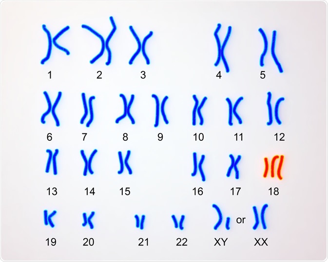
- Edward’s Syndrome
Trisomy 18 – when there are 3 homologous arms of chromosome 18. Sometimes patients can develop something also known as Mosaic Edward’s Syndrome, where a patient has some normal 46 chromosome somatic cells and some trisomy 18 Edward’s Syndrome cells. Symptoms include microcephaly, scrunched fists, arched spine and abnormally shaped chest and umbilical hernia. Common complications include heart defects.
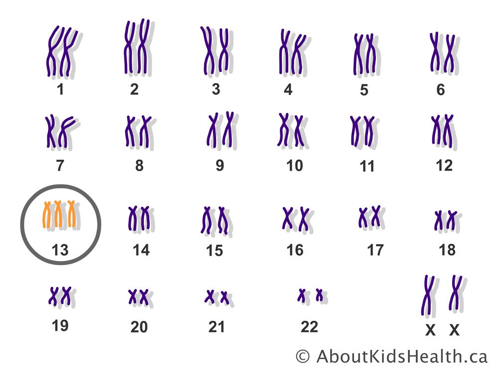
- Patau’s Syndrome
Trisomy 13 – this is where 3 homologous arms of chromosome 13. Effects include polydactyly facial clefting, neural tube defects, and heart defects are also frequent clinical features. Pautau’s syndrome occurs by chance – it is due to random mutations. Usually babies don’t survive after 1 year of life.
There are a lot more stages in the cell cycle than just mitosis. Even mitosis has many sub-stages, which all occur in a certain order for the cell to divide successfully. This allows the cells to accurately divide and make sure no abnormalities occurs during division.

Leave a comment